All of the images below are "thumbnails."
To see the full size pictures, just click on them
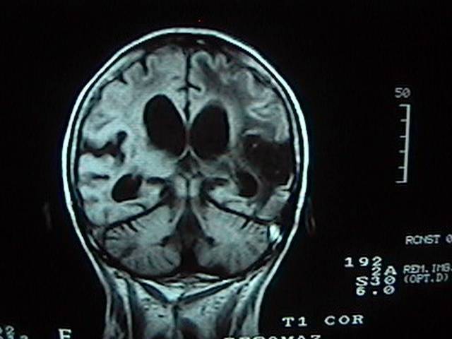 This patient had a sudden loss of her motor functions
(she wasnt able to move her right arms and legs) 2 months
before the study. She went thru a slow recovery with lot
physical therapy and drugs. She was recovering some of her
movements but suddenly all the improvement stop. We
performed an MRI that showed the changes expected for a
lesion of that time (2 months old) but also showed and
increase in the size of the ventricular system( where the
Cerebrospinal fluid or CSF flows) that was causing
hydrocephalus. Due to this finding, the patient went thru
another surgery and had a shunt valve installed, the last
word we had from one of her relatives is that she is again
on recovery.
This patient had a sudden loss of her motor functions
(she wasnt able to move her right arms and legs) 2 months
before the study. She went thru a slow recovery with lot
physical therapy and drugs. She was recovering some of her
movements but suddenly all the improvement stop. We
performed an MRI that showed the changes expected for a
lesion of that time (2 months old) but also showed and
increase in the size of the ventricular system( where the
Cerebrospinal fluid or CSF flows) that was causing
hydrocephalus. Due to this finding, the patient went thru
another surgery and had a shunt valve installed, the last
word we had from one of her relatives is that she is again
on recovery.
The official report included this: T 1 coronal SE (spin echo) sequence that shows an area of infarction in the left parietal lobe. Also enlargement of the ventricular system is observed.
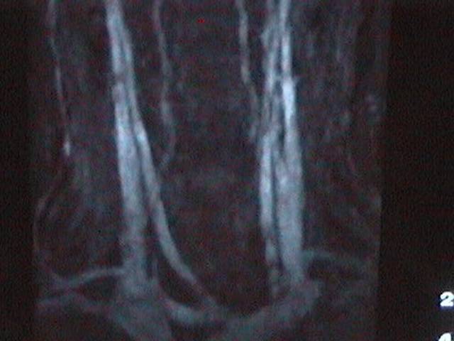 This patient had syncopes every other day...he is a
50 year old male without any important background...MRA
(Magnetic Resonance Angiography) was performed due to the
fact that he has a
short neck and because of that was a bad candidate to check
the entire carotid and vertebral arteries. So, MRA was chosen. it
turn out to have a normal appearance. A Holter (24 hours monitoring
of the heart rythm) disclosed the cause...an unusual arrytmia was
the reason of the sudden loss of blood supply to the brain.
This patient had syncopes every other day...he is a
50 year old male without any important background...MRA
(Magnetic Resonance Angiography) was performed due to the
fact that he has a
short neck and because of that was a bad candidate to check
the entire carotid and vertebral arteries. So, MRA was chosen. it
turn out to have a normal appearance. A Holter (24 hours monitoring
of the heart rythm) disclosed the cause...an unusual arrytmia was
the reason of the sudden loss of blood supply to the brain.
The official report included this: GE (gradient echo) refocused sequence that shows normal blood flow from the carotid and vertebral arteries. MRA (Magnetic Resonance Angiography) DOES NOT USE CONTRAST INJECTION to obtain this images since the flow of blood (with its water component) gives enough signal to display the vessels. Is an extraordinary and non-invasive method to evaluate medium and big vessels of the body.
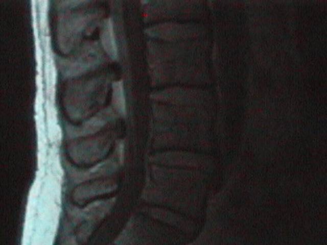 A 39 year old male came due to lower back pain after
moving stuff from his home to a depot. MRI shows a bulging
disk, that means that there is no herniation of the diskal
content. This is very important since the managment does not
require surgery. Only an MRI can tell with such accuracy the
status of the disks, vertebral bodies, ligaments and nerve
roots.
A 39 year old male came due to lower back pain after
moving stuff from his home to a depot. MRI shows a bulging
disk, that means that there is no herniation of the diskal
content. This is very important since the managment does not
require surgery. Only an MRI can tell with such accuracy the
status of the disks, vertebral bodies, ligaments and nerve
roots.
The official report included this: T1 sagital SE (spin echo) sequence of the spine shows a "bulging" disk at L4-L5 level. The vertebral bodies, disk spaces and spine area clearlya observed.
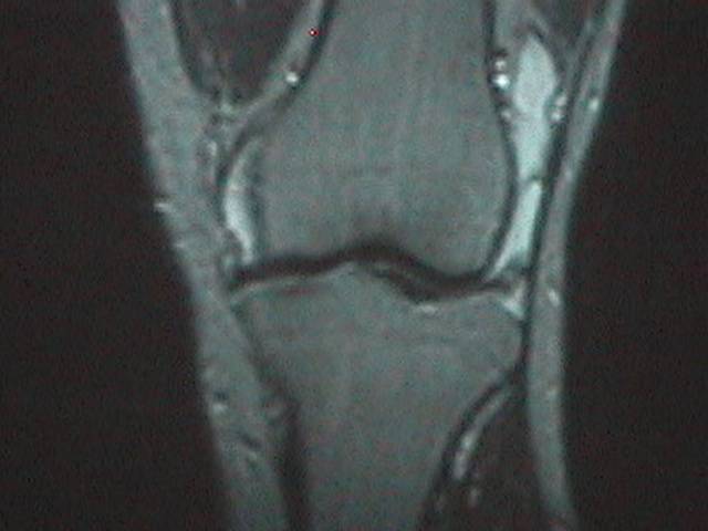 A 30 year old male that after a soccer game came with
swelling of the knee. A meniscal tear was suspected. The MRI
confirmed the lesion and also showed important swelling
within the knee. The appearance of any structure is easily
disclosed in MRI. Here you can actually
see the bones, ligaments, soft tissues and the fluid
collections that appears bright and at surrounds the knee.
A 30 year old male that after a soccer game came with
swelling of the knee. A meniscal tear was suspected. The MRI
confirmed the lesion and also showed important swelling
within the knee. The appearance of any structure is easily
disclosed in MRI. Here you can actually
see the bones, ligaments, soft tissues and the fluid
collections that appears bright and at surrounds the knee.
The official report included this: T2 coronal Se (spin echo) sequence of the knee. The bright (white) rounded images that surround the knee is fluid related to synovitis or inflamation of the bursaes of the knee in a patient with a sport-related injury.
Save the whales. Collect the whole set
Alan Cundall
Sitemap
Go up to Resomaz, Mazatlan's Imaging Center Go up to Some Local Businesses you might Consider Go up to What Others Say Go up to General Information on Mazatlan Go up to Home Page of Nadine Loves Henry
Go back to A Tour of Resomaz -2- Continue with Special Deals for Tourists